
EDTA Chelation Dissolves the Artificial Intelligence Magnetic Hydrogel Weapon
Patent Review Shows Hopeful Data
I have been advocating for months that EDTA Chelation as a treatment solution for the C19 injected, as it effectively removes metals, and can detoxify the body even from Graphene. As a certified Chelation practitioner, it is evident to me that EDTA is needed for the environmental bombardment of this AI parasitic life form, that we are exposed to via C19 injections, chemtrails, vax shedding, food, cosmetics, medication and many other avenues.
We now know that the self-assembly nanotechnology that grows to micrometer size grows out of the lipid nanoparticles. Karen Kingston found the patents:
My colleague Dr. Shimon Yanowitz showed that the metal nanoparticles self assemble out of the Liposomes.
The only thing that is not consistent is the scale. We see micrometer size and they speak of nanometer size. This shows that the self assembly already is active upon thawing of the C19 vials.
The patent states:
CELL-FRIENDLY INVERSE OPAL HYDROGELS FOR CELL ENCAPSULATION, DRUG AND PROTEIN DELIVERY, AND FUNCTIONAL NANOPARTICLE
FIELD OF THE INVENTION
The invention relates to polymer scaffolds for cell-based tissue engineering.
BACKGROUND
Tissue engineering is an approach for regeneration, replacement, and improvement of the functions of damaged tissues by manipulating materials according to the specific structure or function of the desired tissues. Porous and biodegradable polymer scaffolds, e.g., three dimensionally interconnected scaffolds, are utilized as a structural supporting matrix or as a cell adhesive substrate for cell-based tissue engineering. A highly open porous structure with interconnected pores is required to achieve sufficient cell seeding and migration within the scaffold, as well as to facilitate mass transfer of nutrients, oxygen, and metabolite waste for sequential proliferation and differentiation of large quantity of cells. Current approaches to generate porous networks in polymer scaffolds include gas foaming, salt leaching, and freeze drying; however, the limitations of those processes include irregular pore sizes, shapes, and structures, as well as limited interconnectivity. As such, there is a pressing need in the art to develop improved structured polymer scaffolds with interconnected pores.
SUMMARY OF THE INVENTION
The invention described herein provides the fabrication of cell- friendly inverse opal hydrogels that also allow cell-encapsulation in the hydrogel matrix. An inverse opal hydrogel scaffold device comprising a polymer matrix and a sacrificial porogen in which the porogen comprises an ionically-crosslinked polymer, a thermosensitive polymer, a thermoresponsive polymer, a pH-sensitive polymer, or a photocleavable polymer. The polymer matrix is made of a durable polymer relative to the sacrificial porogen such that the polymer matrix withstands physical or chemical changes that cause porogen sacrifice. For example, polymer matrix is covalently crosslinked, withstands a change (e.g., increase) in temperature, withstands a pH change (e.g., decrease) or change in ionic strength or composition (e.g., contact with a divalent cation chelator), or withstands exposure to light (e.g., UV light).
For tissue engineering and cell scaffold applications, the polymer matrix further comprises an isolated cell, e.g., a eukaryotic cell. By "isolated cell" is meant a cell that has been separated from the other cells, components, and/or environment that naturally accompany it. Alternatively, the matrix contains prokaryotic cells such as bacteria. For example, the polymer matrix is crosslinked and comprises an isolated cell encapsulated in the crosslinked polymer matrix. An exemplary polymer matrix comprises a synthetic polymer such as one that is covalently crosslinked. Examples of polymer matrices include poly(lactide-coglycolide) (PLGA; a copoly lactic acid/glycolic acid polymer), poly(acrylic acid), polyethylene glycol (PEG), poly (vinyl alcohol), or polyphosphazene. The sacrificial porogen comprises an ionically-crosslinked polymer, a thermosensitive polymer, a thermoresponsive polymer, a pH-responsive polymer, or a photo-cleavable polymer. Exemplary polymers for a porogen include alginate, collagen, gelatin, fibrin, agarose, hyaluronic acid, or chitosan as well as thermosensitive polymer such as agarose, gelatin, or collagen, poly(N-isopropylacrylamide), poly(N-ethylacrylamide), poly(N- cyclopropymethacrylamide), poly(N-methyl-N-ethylacrylamide), poly(N- acryloylpyrrolidine) , poly(N-ethy lmethacry lamide) , poly(N-cy clopropylacry lamide) , poly(N-cyclopropylacrylamide), poly(N,N-diethylacrylamide), poly(N- isopropy lacry lamide) , poly(N- inylcaprolactam) , poly(N-n-propy lmethacry lamide) , poly(N-methyl-N-isopropylacrylamide), poly (N-n-propy lacry lamide), poly(N-methyl-N-n- propy lacry lamide), and poly(N-acryloylpiperidine).
Hydrogel (also called aquagel) is a network of polymer chains that are hydrophilic, and are sometimes found as a colloidal gel in which water is the dispersion medium.
Hydrogels are highly absorbent (they can contain over 99% water) natural or synthetic polymers. Hydrogels also possess a degree of flexibility very similar to natural tissue, due to their significant water content. Hydrogel shaped as an inverted opal exhibits much higher swelling ratios, and its swelling kinetics is an order of magnitude faster as well. The engineered scaffolds (i.e., inverse opal hydrogels) described herein possess desirable mechanical and optical properties that can facilitate tissue regeneration while allowing for continuous high-resolution optical monitoring of cell proliferation and cell-cell interaction within the scaffold.
Methods of producing an inverse opal hydrogel with open, interconnected pores are carried out by compressing a plurality of template porogen particles into a mold, and subsequently adding a composition comprising a polymer solution and a plurality of cells to the interstitial space between template porogen particles in the mold to polymerize the template porogen particles. The template porogen particles are removed from the mold, thereby producing an inverse opal hydrogel with open, interconnected pores, wherein the cells are encapsulated in the inverse opal hydrogel. The template porogen particles are removed without using toxic organic solvents or lyophilization. For example, thermosensitive hydrogel beads are removed by controlling the temperature to change the solid phase of the beads. The template porogen particle is an ionically crosslinked polymer, a thermosensitive polymer, a thermoresponsive polymer, a pH-responsive polymer, or a photo- cleavable polymer.
For example, the ionically crosslinked polymer is alginate. The ionically crosslinked polymer is removed by adding a metal-chelating agent selected from the group consisting of citric acid, ethylenediamine, ethylenediaminetetraacetic acid (EDTA), diethylenetriaminepentaacetic acid (DTP A), and N,N-bis(carboxymethyl)glycine (NTA).
Suitable thermosensitive polymers include agarose, gelatin, and collagen. The thermosensitive polymer is removed by increasing the temperature of the polymer, thereby altering the phase of the polymer to liquid phase. Examples of photocleavable polymers include chromophore-based crosslinkers for photodegradable hydrogels, (4-vinylpyridine) (P4VP) and poly (methylmethacrylate). Methods of producing an inverse opal hydrogel with open, interconnected pores are carried out by compressing a plurality of template porogen particles into a mold, adding a composition comprising a polymer solution and an agent to the interstitial space between template porogen particles in the mold to polymerize the template porogen particles, and removing the template porogen particles, thereby producing an inverse opal hydrogel with open, interconnected pores. The agent is selected from the group consisting of a drug, a nanoparticle (e.g., magnetic nanoparticles or gold nanoparticles), a growth factor (e.g., vascular endothelial growth factor (VEGF), platelet derived growth factor (PDGF), brain derived neurotrophic factor (BDNF), epidermal growth factor (EGF), or fibroblast growth factor (FGF)), a cytokine (e.g., interferon gamma (IFN-γ), erythropoietin (EPO), thrombopoietin (TPO), interleukin-1 (IL-1), IL-4), a chemokine (e.g., a CC chemokine, a CXC chemokine, a C chemokine, or a CX3C chemokine), a hormone (e.g., insulin, growth hormone, vasopressin, testosterone, or Cortisol), a protein, a nucleic acid, or a small molecule. In one example, nanoparticles are encapsulated within the hydrogel matrix, and cells are dispersed within the open-interconnected pores.
Example 1 : Alginate Beads as a Sacrificial Template
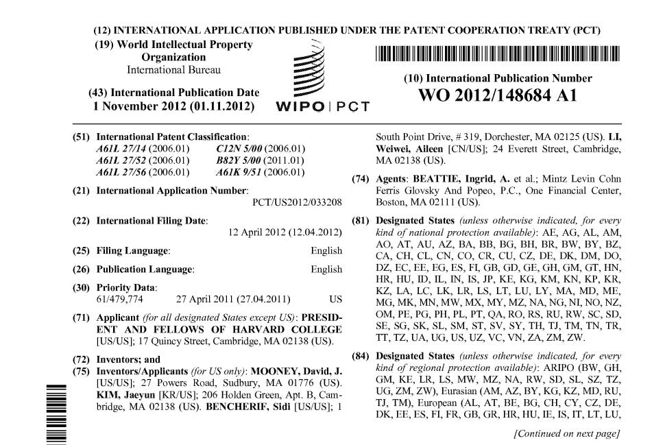
Described herein is an example of the fabrication of a cell- friendly inverse opal hydrogel. Alginate beads, formed using Ca2+-crosslinking were used as the porogen, and 50 mM EDTA solution, a metal chelating agent, was used as the template removal solution. To evaluate if EDTA can dissolve the alginate beads efficiently, three different sized alginate beads were prepared using 2% alginate solution in 100 mM Ca2+ solution (Figure 3, upper row). Rhodamine-labeled bovine serum albumin (BSA) was encapsulated in alginate beads to visualize the beads and their dissolution. The resulting alginate beads were incubated in 50 mM EDTA solution under shaking. After 20 min, all alginate beads dissolved in EDTA solution and lost their spherical morphology, and this resulted in a pink solution due to released rhodamine-labeled BSA from alginate beads (Figure 3, lower row). This demonstrates that the alginate beads were used as sacrificial template by using EDTA as leaching solution.
Figure 3 is a series of photomicrographs demonstrating that ethylenediaminetetraacetic acid (EDTA) efficiently dissolves alginate beads in IOHs.
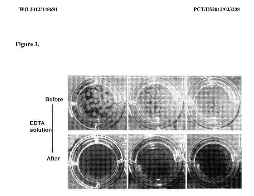
Figure 5 a is a series of photomicrographs showing cell viability after treatment with EDTA for up to 3 hours. Figure 5b is a bar chart showing about 98% viability of cells after 3 hour incubation in EDTA.
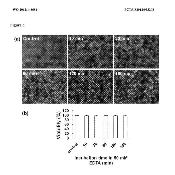
Figure 6a is a series of photomicrographs showing cell viability after treatment with EDTA for up to 7 days. Figure 6b is a bar chart demonstrating the proliferation of cells encapsulated in IOHs.
Example 3: EDTA is Non-Toxic to Cells Encapsulated in IOHs
To evaluate if incubation in EDTA solution is toxic to cells, cell viability was checked after incubation in 50 mM EDTA solution. Mouse mesenchymal stem cells (MSCs) were cultured in a flask, and incubated in a 50 mM EDTA solution for 10, 30, 60, 120, or 180 min. Subsequently, the viability of cells was measured with a live/dead cell assay by using calcein AM and ethidium homodimer-1. Although the cell morphology changed to a round shape, the representative fluorescent images of the live/dead assay showed that there was no significant toxicity of the EDTA solution to the cells for up to 3 h incubation(Figure 5a). Quantitative analysis also showed -98% viability even after 3h incubation in EDTA solution (Figure 5b). Based on these observations, incubation of the gels in 50 mM EDTA solution for up to 3 h to remove alginate beads was determined as a nontoxic process to cells encapsulated in IOHs.
Ionically crosslinked hydrogel beads (e.g., alginate) are removed by using various metal-chelating agents including citric acid, ethylenediamine, ethylenediammetetraacetic acid (EDTA), diethylenetriaminepentaacetic acid (DTP A), N,N- bis(carboxymethyl)glycine (NTA), etc. The chelating agents bind with metal ions used as the crosslinker of templating beads, which results in the generation of pores via the dissociation of metal ions and polymers forming beads.
For example, various chemical drugs including small molecules and functional proteins (growth factors, cytokines, chemokines, hormones, etc) are mixed with the polymer precursor solution to be added into the template beads in the mold. After polymerization of the polymer precursors and removal of the template beads, the encapsulated molecules are released slowly. The release profiles depend on crosslinking density, the affinity of molecules to the polymer chain, the size of molecules, etc. In this context, the inverse opal hydrogels are used as delivery systems for the cells encapsulated in the hydrogel or the cells outside the hydrogel.
The methods described herein also encapsulate functional nanoparticles to actuate the porous hydrogel systems to release cells, drugs, proteins, and growth factors on demand. The nanoparticles are encapsulated in the hydrogel in a similar manner in which cells and drugs are encapsulated. Specifically, the functional nanoparticles are mixed with polymer precursors and added into the template beads in the mold. For example, magnetic nanoparticles or gold nanoparticles are encapsulated in the polymer matrix and the resulting porous hydrogels are responsive to an external magnetic field or light, respectively. The guest molecules are released upon detection of the external stimulus. In the case of magnetic nanoparticles, the external magnetic force modulates the volume of pores in the inverse opal hydrogel due to its high porosity. The guest molecules encapsulated or seeded in the inverse opal hydrogels are released by the mechanical forces via convection. In addition, both magnetic nanoparticles and gold nanoparticles are used as hyperthermic moieties. Magnetic nanoparticles and gold nanoparticles generate heat by alternating magnetic fields and irradiation with lasers, respectively. Thus, both magnetic and gold nanoparticles allow thermal motion of the polymer matrix and the encapsulated guest molecules, which accelerates the release rate of guest molecules.
Summary and next steps:
This patent shows that the Hydrogel can be dissolved with EDTA Chelation. The metal nanoparticles that are in the Hydrogel to change its magnetic properties and regulate the release of guest molecules, for example toxins or other substances, can also be removed from the body by EDTA. It is my suspicion that the ribbons we see in live blood analysis of C19 injected people as well as in the un injected via shedding and environmental exposure are made of this hydrogel that also can have carbon nanotubes in various forms associated with it as well as metallic nanoparticles. WE MUST TEST THE LARGE CLOTS FOUND IN THE DECEASED INJECTED TO SEE IF THIS IS AI HYDROGEL AS KAREN KINGSTON SUGGESTS. My suspicion is that she is absolutely right. Fibrin can be one of the guest molecules and the metal particles of Aluminum and Tin that Mike Adams found in his analysis, also suggest what is found in this patent - a hydrogel polymer with metal nanoparticle enclosures.
EDTA Chelation should be utilized in all vax injury treatment protocols and prophylactically for the injected and un injected alike - to treat this unprecedented attack of diabolical Artificial Intelligence parasitic life form weapon upon humanity.
All of us medical practitioners who do Live blood analysis need to look at before and after samples to see if dissolution of ribbons occurs and how many treatment it takes to clear the blood. Routine repetition of EDTA Chelation IV Therapy should be done to prevent re accumulation of this artificial life form in the body, in particular since the exposure now is coming from literally everywhere.
If you have not yet seen this brilliant interview of Karen Kinston with Maria Zeee, please do so right now :
STOP THE SHOTS. STOP GEOENGINEERING. STOP 5G. STOP POISENING OUR FOOD AND WATER SUPPLY. STOP POISENING THE BODY WITH BIG PHARMA TOXINS.
Special Thanks to Karen Kingston for her brilliant work in finding these patents.


No comments:
Post a Comment