Environmental Filaments UV Light Fluorescence Darkfield Microscopy
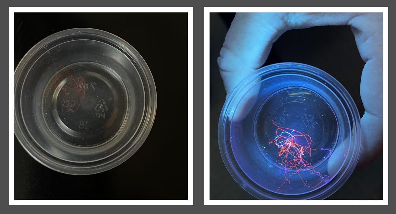
Image: Environmental filaments collected in regular light and under UV light
I was visited by Dr. Justin Coy, a former Defense Department Contractor who has been following and validating my research. He brought me an environmental filament sample and a UV flashlight - 365nm. In this post, I am documenting the darkfield microscopy of these filaments and experiments with UV light. He was suspecting Luciferase to be present in the filaments and asked me to take a look. From my research there are metal nanoparticles in the filaments and they can cause fluorescence. Luciferase is used in in molecular biology that uses the luciferase enzyme and a substrate (such as luciferin) to study gene regulation at the level of transcription. I do not think that is the mechanism of the fluorescence of the polymers as other mechanisms using metals have been described in the literature and since Clifford Carnicoms analysis showed huge amounts of metals in the filaments that could be plausible.
There have been developments of bright orange proteins fused with Luciferase in biological systems - this remains a question for further research and discovery.
Novel NanoLuc substrates enable bright two-population bioluminescence imaging in animals
We know that embedded Quantum Dot technology can make filaments emit different light and filaments found in the blood have been shown to have bifringence. We also know that UV light can be used as an energy source by nano sensors which can embed themselves in the self assembly polymers. Karl C has done some remarkable microscopy research showing this unusual light emission which I posted here: Extraordinary Microscopy Of Self Assembly Nanotechnology - A Request For Funding Help For Karl C
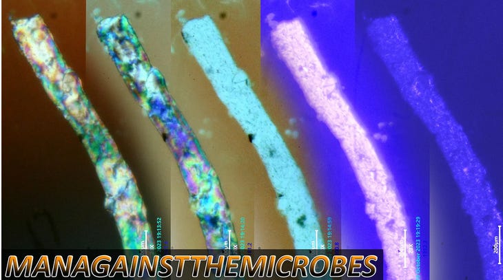
Here are different images of the filaments analyzed by me observing how they change with normal light and then UV light:
Image: Darkfield Microscopy: UV light off left, UV light on right
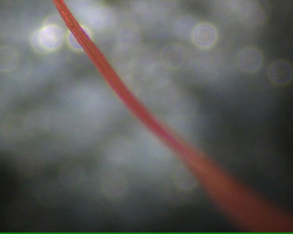
Image above: normal light
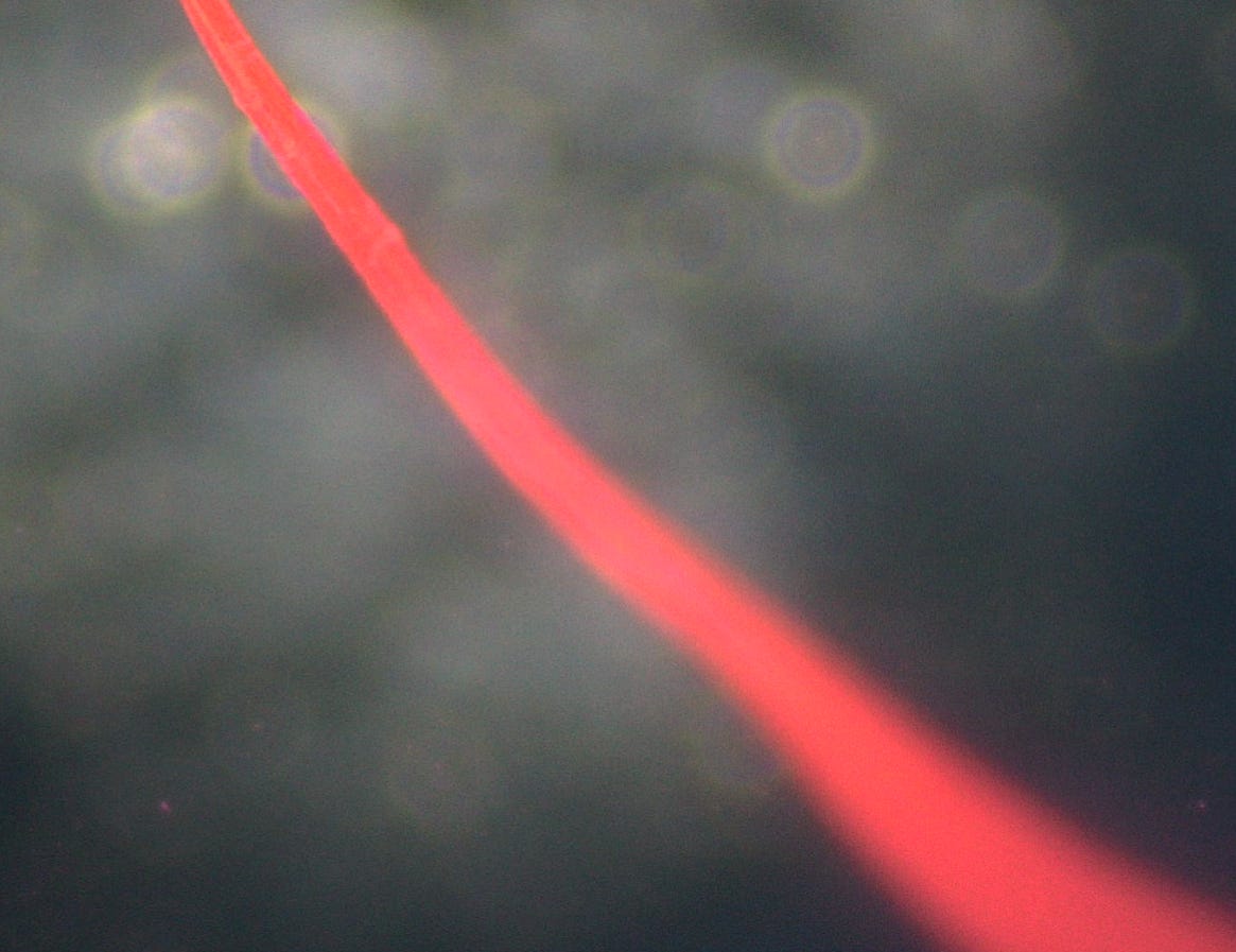
Image: UV light
I then wanted to see if different aspects of the filament react differently to UV light, and they appear to. Some areas are more luminescent then others.
Image: UV light on, both pictures.
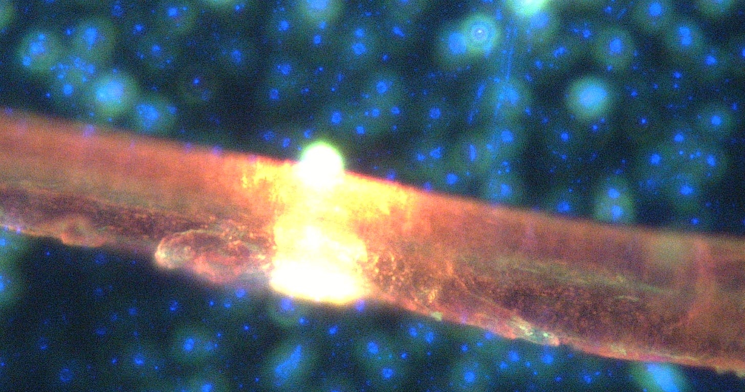
Below you can see a closer view of the filament under UV light, and there being very specific region that react to the UV light more:
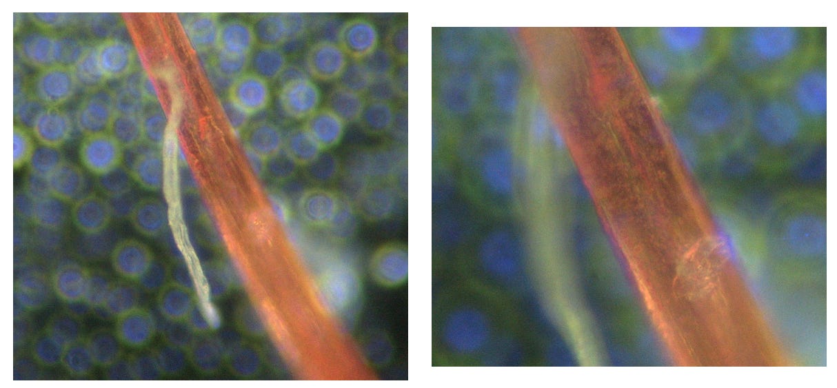
Off the orange appearing filament a white one came forth. Magnification of 2000x on the right shows a central cavitation of the filament
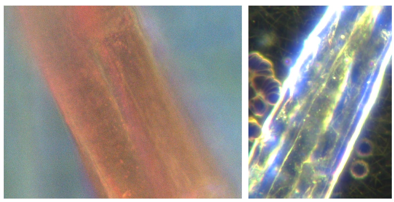
Below you can see the orange environmental filament compared to a "self assembly nanotechnology hydrogel” filament from shedding in C19 unvaccinated blood with many visible Quantum Dot like structures seen embedded. The filament composition look the same except the colors differ.
Here are different areas of the filament that have enormous glow under UV light, magnification 2000x:
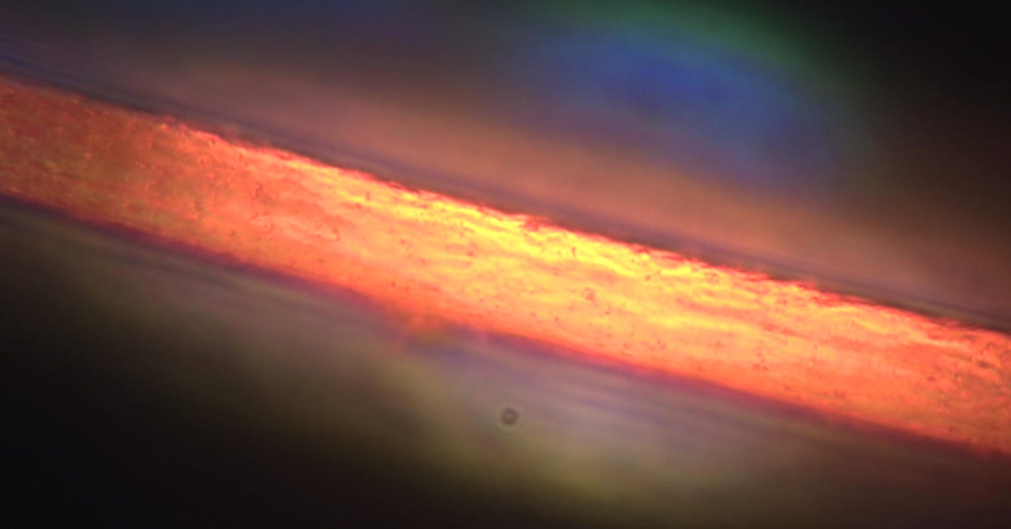
Here is an area of the filament with UV light:
Same without UV light:
Here are some research articles on fluorescent polymers:
New 'smart' polymer glows brighter when stretched
Spider fossils glow under UV light, a clue to their remarkable preservation
I have been speaking about spider silk which is a polyamide protein and recently did microscopy on an environmental filament found:
This article explains that if metals are introduced the the nanofibers, florescence can be achieved:
Optical fluorescent spider silk electrospun nanofibers with embedded cerium oxide nanoparticles
The work demonstrates an electrospun nanocomposite of recombinant spider silk protein (rSSp) nanofibers with embedded cerium oxide (ceria) nanoparticles. RSSP (MaSp1) has been produced, extracted from goat milk, and fabricated into nanofibers using an electrospinning process. The resulting electrospun nanofibers have a mean diameter of ∼50 nm. Furthermore, ceria nanoparticles of mean diameter 10 nm were added in the spinning dope to be embedded within the generated nanofibers. These nanoparticles show certain optical activity due to optical trivaliant cerium ions, associated with formed oxygen vacancies. The formed nanocomposite shows promising mechanical properties such as the Young's modulus, elasticity (or elongation at break), and toughness. In addition, the electrospun mat becomes fluorescent with 520-nm emission upon exposure to UV light, due to excitation of the optically active ceria nanoparticles. Also, the formed nanocomposite shows a decay of its electric resistance over time upon exposure to cyclic loads at different humidity conditions. The synthesized nanocomposite can be utilized in different biomedical, textile, and sensing applications.
We do know that these polymers are used for transhumanist surveillance and synthetic biology. Here they used spider silk as an inspiration. Note how they describe that these polymers can wrap around nerves, muscles and hearts and be the next generation tissue electronic interface:
Polymer films inspired by spider silk connect biological tissues and electronic devices
Linking biological tissues with electronic devices is challenging owing to the softness of tissues and their arbitrary shapes and sizes. An innovative water-responsive, supercontractile polymer film, inspired by spider silk, allows the construction of soft, stretchable and shape-adaptive tissue–electronic interfaces.
We designed water-responsive supercontractile polymer films composed of poly(ethylene oxide) and poly(ethylene glycol)-α-cyclodextrin inclusion complex, which are initially dry, flexible and stable under ambient conditions, contract by more than 50% of their original length within seconds (about 30% per second) after wetting and become soft (about 100 kPa) and stretchable (around 600%) hydrogel thin films thereafter. This supercontraction is attributed to the aligned microporous hierarchical structures of the films, which also facilitate electronic integration. We used this film to fabricate shape-adaptive electrode arrays that simplify the implantation procedure through supercontraction and conformally wrap around nerves, muscles and hearts of different sizes when wetted for in vivo nerve stimulation and electrophysiological signal recording. This study demonstrates that this water-responsive material can play an important part in shaping the next-generation tissue–electronics interfaces as well as broadening the biomedical application of shape-adaptive materials.
Here is video of the microscopy UV light on in both videos:
UV light on playing with the focus:
I took a blood sample and applied the UV light to see what happens to the micro robots. As in my experiments with the 450nm cold laser, the robots are quite happy and seem to absorb the extra energy - if you look at the robot its light emission intensifies, and that is consistent with the WBAN article I just posted that light is an energy source for the biosensors. Energy Harvesting From The Human Body By Wireless Body Area Network - A Cause For The Electrical Conductivity Loss in Human Blood?






No comments:
Post a Comment